ANATOMY OF AN IXODID TICK
General anatomy
Generalised Ixodid female
Generalised Ixodid male
Capitulum
Chelicerae
Limbs
Viscera
General Structure of Ixodid Ticks
Ticks are closely related in general body form to parasitic mites.The hard (ixodid) ticks have the following general characteristics- 1. a teardrop body shape (argasids are oval), with mouthparts forward/anterior (argasids ventral) 2. adults have a genital aperture nearly between coxa III (argasids between coxa I), a pair of spiracles posterolateral to coxa IV (argasids posterolateral to coxa III), eyes absent or present (immature or adult ticks) (argasids eyes usually absent), well developed pulvilli (argasids pulvilli absent or poorly developed exc in a few larvae), four well-marked plates (ventral, anal and two lateral), posterior margins scallop-patterned festoons present or absent (argasids festoons absent). 3. larval stages also possess an anterior scutum and the entire dorsal capitulum can be seen from above (argasid larvae have mid-dorsal oval scutal shields and only the mouthparts are usually visible from above) (Cupp, EW, 1991).
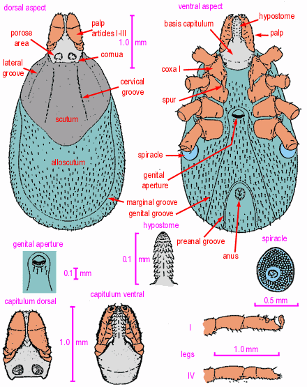
Sexual dimorphism is pronounced. Ticks of this family possess a hard, chitinous shield or scutum which extends over the whole dorsal surface of the male but covers only a small portion behind the head in the larva, nymph and adult female. In the male the body is incapable of becoming greatly enlarged (probably because it is constrained by the scutum). In the female the body is capable of considerable enlargement. The scutum (literally, the shield) is sometimes referred to as the face but this is a misleading term.
The general body of the tick may be divided into capitulum (mouthparts) and idiosoma (body). The idiosoma in turn is comprised of the podosoma (limbs) plus the opisthosoma (torso).
The scutum has bilateral cervical and lateral grooves, varying
in depth and length in different species. The body of the female
may have a pair of lateral "marginal grooves" behind
the scutum, while postero-lateral and median grooves are usually
present on the dorsum in both sexes. The posterior border of the
body is frequently notched or indented, forming the "festoons"
which generally number eleven. The genital opening is a ventral
transverse slit in front of the middle, the anus being posterior.
The male may have ventral plates. Ornate ticks may have coloured,
enamel-like areas on the body, inornate ticks have not.
Generalised female Ixodid tick
(as per Ixodes cookei); based on Sonenshine, DE: Biology of Ticks, 2 volumes: Oxford University Press, New York, Oxford, 1991:
Generalised male Ixodid tick
(as per Ixodes cookei); based on Sonenshine, DE: Biology of Ticks, 2 volumes: Oxford University Press, New York, Oxford, 1991:
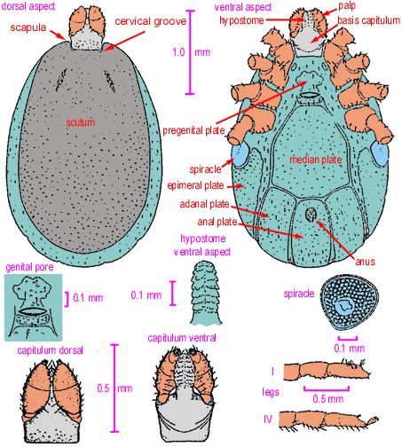
Spiracular plates are located posterior, somewhat lateral to coxa IV.
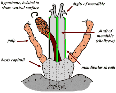
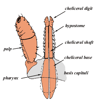
The capitulum of an Ixodid tick
Also called the "rostrum", "the head", or "false head"and the "snout" the capitulum is the moveable anterior extension of the body and includes both the palps and the mouth parts (basis capituli, chelicerae and hypostome).
 .
.
An Ixodid capitulum based on Amblyomma spp, dorsal aspect (adapted from: Soulsby, E J L; Helminths, Arthropods and Protozoa of Domesticated Animals; Bailliere Tindall and Cassell, London, 6th ed, 1968.).
The capitulum is articulated with the body via a cavity (the emargination in ixodids and the camerstome in argasids), and normally lies in the same plane as the body. It is connected by a soft articulation membrane that allows the capitulum to be flexed (ventrally) or extended (returned to the horizontal axis).
The basis capituli shows two dorsal porose areas in the female. The mouth parts are anterior and well visible from the dorsal aspect.
capitulum of Ixodes holocyclus nymph, dorsal aspect, based on Nuttall, 1908
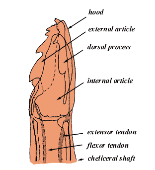
Chelicerae
The chelicerae lie dorsal to the hypostome and support the cheliceral digits which cut into the skin of the host.
Limbs of an Ixodid Tick
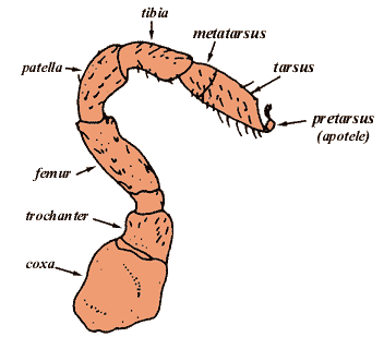
The first pair of limbs forms the chelicerae (mandibles) for cutting into the skin of the host, the second pair of limbs forms the palps which position the capitulum for feeding, the third to sixth pairs of limbs are the actual legs used for walking whilst on and off the host. The four pairs of legs are divided into 6 segments- coxa, trochanter, femur, tibia, metatarsus and tarsus. The coxae are situated on the ventral side of the body and have very limted movement. The other segments are highly flexible but are mainly used by flexing and extending the joints. The may be folded against the ventral body wall for protection. The segments are connected by a soft articulation cuticle. The tarsus of each leg bears an apotele (=pre-tarsus), including the claws and and the pulvillus. There is a complex sensory apparatus (=Hallers organ) located on the dorsal surface of the tarsus of leg I. This is very important, as it is the primary organ for determining host location, and detecting host odors and pheromones etc.
Viscera of a representative male Ixodid Tick
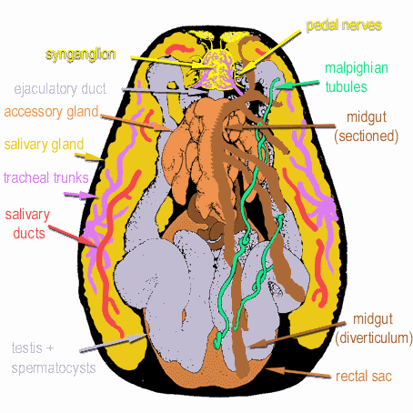
Relative positions of the internal organs of a representative male ixodid tick. The left hand side has had the midgut removed along the midline as indicated by the jagged edge. The midgut diverticula overly most of the other organs. The synganglion represents the "brain" of the tick. The tracheal trunks converge to form the spiracle on the tick's ventrolateral surface. Adapted by NF from drawings by Ms M Bloomfield, Norfolk, Virginia; in Sonenshine DE (ed), Biology of Ticks, Oxford University Press, 1991.
The Paralysis Tick of Australia - Home
E-mail Us to report a broken link!