|
MEGAOESOPHAGUS
IN DOGS
|
Dogs may be born with esophagus or they may develop it later in life.
It is commonly seen with other disorders such as myasthenia gravis
and peripheral neuropathies. Megaesophagus is the result of an esophageal motor disorder causing hypomotility. The congenital form is noticed after weaning and is hereditary. Onset of the acquired form may be at any age and has been associated with viral encephalitis, lead poisoning, myasthenia gravis, immune mediated polyneuritis, SLE, polymyositis, hypothyroidism, hypoadrenocorticism, myotonia, trauma, tick paralysis, various neuropathies and many other causes. There is often secondary pneumonia. |
|
|
|
|
|

|
| Unless an underlying cause can be found, there is no cure for
megaesophagus.
What is it?
Megaesophagus refers to a syndrome in which the esophagus becomes weak and flaccid, and subsequently becomes much larger than normal, hence the term megaesophagus. This occurs because the muscles of the esophagus lose tone. Once this occurs, the esophagus does not propel ingested food, air, and water into the stomach. Rather, these items remain in the esophagus for prolonged periods of time.
This syndrome is much more common in dogs than cats, and can occur in dogs of any age. There are many causes of megaesophagus, but the consequences tend to be similar regardless of cause. Affected pets usually regurgitate fluid or food. Regurgitation is much like vomiting, except that vomiting involves forceful ejection of material from the stomach and intestine, whereas regurgitation involves the more passive emptying of material from the esophagus or back of the mouth. Regurgitation related to megaesophagus may occur soon after eating or hours later. Dogs may or may not lose weight, depending on how much food ultimately reaches the stomach.
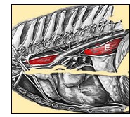 Pertinent Anatomy Pertinent Anatomy
The esophagus is a long muscular tube that connects the mouth to the stomach (illustration right - esophagus as it passes through the chest cavity - red structure
labelled E).
The purpose of the esophagus is to transport swallowed food and water to the stomach. To do this, the esophagus normally uses a squeezing movement behind the food (peristalsis) to propel the material into the stomach within seconds after it is swallowed. After swallowing, the normal esophagus is empty and resembles a collapsed hose.
Diagnosis
Megaesophagus is diagnosed by taking radiographs (x-rays) of the chest. Occasionally contrast studies (feeding food with barium) are needed to outline the esophagus. Constant motion radiographic studies (fluoroscopy) looking at esophageal motility may be required in patients that have inconclusive thoracic (chest) radiographs. It is important to obtain these studies because there are other problems that can cause clinical signs similar to megaesophagus but require very different therapy.
Because of the potentially devastating side effects of megaesophagus, it is wise to look for an underlying cause. Frequently, the underlying cause is found in only very few patients.
|
|
Treatment
Treatment consists of trying to help the food get from the mouth into the stomach. If food does not remain in the esophagus, it cannot be regurgitated and subsequently aspirated into the trachea or lungs. Dry, canned, and gruel diets should be tried to find the one best handled by your pet. Feeding several small meals a day is generally preferred over feeding 1-2 larger meals. Medications may be given to help decrease gastroesophageal reflux, the movement of stomach acid into the
esophagus.
For patients severely affected, a tube can be placed into the stomach through the body wall (gastrostomy tube). This allows the dog to receive food and water without having to go through the esophagus. This feeding technique does not eliminate the possibility of aspiration, as the dog is still swallows saliva, but may help to diminish it. These feeding tubes can remain in place for long periods of time; however, and depending on the type used, it may need to be replaced periodically.
Prognosis
Unless an underlying cause can be found, there is no cure for megaesophagus. In some patients, the regurgitation of food will become worse over time, whereas in others there is no change in the frequency of regurgitation. In those patients with progressive worsening of disease, weight loss becomes a major problem.
The most significant crisis patients face with megaesophagus is the inadvertent transfer of food, water, and saliva into the trachea (windpipe) and lungs. This is called aspiration and can lead to pneumonia. In some instances the dog will show signs of aspiration pneumonia (i.e. cough, labored breathing, fever) despite the owners never having seen evidence of regurgitation. This is because the dog may regurgitate the material into its mouth and then swallow it or inhale it without ever having the material leave its mouth. If only small amounts of material are aspirated into the trachea, cough will be the most obvious problem. This cough may be moist or dry. If larger amounts are inhaled and the material reaches the lungs, severe pneumonia may result, causing fever and labored breathing. Nasal discharge can occur when material is pushed into the back of the nose during regurgitation.
The major cause of death in patients with megaesophagus is aspiration pneumonia. If large amounts of material are aspirated and reach the lungs, the dog can develop sudden, severe pneumonia and the can die form asphyxiation (lack of oxygen). Such a sudden death can occur at any time, even if the dog has not been regurgitating for several weeks or months.
Michigan Veterinary Specialists
Web Site |
|
|
Breeds Affected with Megaesophagus
- Shar Pei
- German Shepherd
- Great Dane
- Greyhound
- Irish Setter
- Miniature Schnauzer
- Wire-Haired Fox Terrier
|
|
|
The causes of Megaesophagus are diverse and include:
- Foreign body in the esophagus
- Tumor (cancerous or noncancerous) in or around the esophagus
- Narrowing (stricture) of the esophagus
- Congenital abnormalities (present at birth)
- Diseases of the muscle and nervous systems, such as myasthenia gravis or lupus
- Infections of the nervous system
- Botulism
- Nerve damage
- Trauma to the brain or spinal cord
- Inflammation of the esophagus
- Hypothyroidism (inadequate production of thyroid hormone by the thyroid gland)
- Poisoning, such as lead poisoning
- Hypoadrenocorticism (Addison's disease, inadequate production of adrenal hormones by the adrenal glands)
- Hereditary condition in Wire Fox Terriers and Miniature
Schnauzers
|
|
|
Signs of Megaesophagus
- Abnormal breath odor
- Abnormal breathing sounds of the upper
airway
- Abnormal lung or pleural sounds
- Anorexia
- Coughing
- Dehydration
- Dullness
- Dysphagia
- Dyspnea
- Erythema
- Redness of
pharynx
- Excessive salivation
- Fever, Gagging
- Generalized weakness
- Increased respiratory rate
- Lack of growth or weight gain
- Mucoid nasal discharge
- Polyphagia
- Purulent nasal discharge
- Sneezing, Tetraparesis
- Underweight, poor condition, thin, emaciated, unthriftiness, ill
thrift
- Vomiting or regurgitation
- Weight loss
|
|
| LINKS
TO RELATED ARTICLES |
|
|
|
|
|
| |
|
|
|